Cervical Spine X-Ray: Medical Imaging of the Human Neck Spine
Description
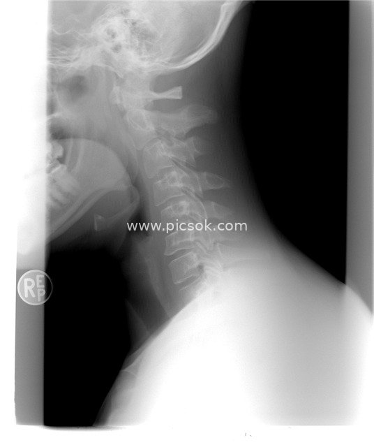
This black-and-white grayscale X-ray image accurately captures the skeletal outline of the human cervical spine. On the left side, parts of the skull, mandible, and oral cavity teeth are faintly visible, while the dark background on the right highlights the clear structure of the neck spine: neatly arranged vertebral bodies, distinct intervertebral spaces, and a softly presented natural physiological curvature of the cervical spine. From a professional medical perspective, the image intuitively displays the internal architecture of the neck spine. The strong contrast between gray-white and black ensures that skeletal details are clearly distinguishable. With a rigorous and professional overall atmosphere, it serves as high-quality visual material for analyzing cervical spine health in orthopedic diagnosis and medical education, helping physicians or learners intuitively understand the anatomical structure and morphological characteristics of the neck spine.